Love Is in the Air...and the Eyes

We have all heard the term ”Love is in the Air,” but did you know that love can be wonderfully expressed through the eyes?  Certain chemicals (or endorphins) that help produce the emotion of love can be emitted through emotions expressed in the eyes. There are physiological changes in the eyes that occur when love is expressed between two individuals. Love for a romantic partner, a family member, or a favorite pet can all cause the same physical response: the pupil (black part in the center of the eye) dilates.

The size of the pupil can be an indication of emotional responses and messages. According to Scientific American, the autonomic nervous system (ANS), which directs our fight or flight response, causes the pupil to have a quick dilating response. The ANS is also in charge of heart rate and perspiration, and when a person is extremely interested in another person, the pupil has a dilating effect that is slightly less than the pupillary light reflex. This bounce in size is an automated response that gives scientists indication of mood or interest (or love) shown to a person or pet.

Mounting scientific evidence also shows the benefit of looking into the eyes of your pet, especially dogs. Stroking them causes you to become more healthy on all fronts, and a few minutes a day of lovingly looking at the dog and stroking the pet releases serotonin, prolactin, and oxytocin, which are "feel good" hormones.

There are also studies inspired by psychologist Arthur Aron, from over 20 years ago, that show if you stare into someone's eyes for 4 minutes you can fall in love... looking eye to eye allows you to connect with someone new or reminds you why you fell in love with this person in the first place.

Overall health is improved and years can be added to your life when looking at something or someone with love. Your eyes and autonomic nervous system play an intricate role in the expression of love.

Source:

12/07/2012 article of Scientific American entitled:

Why Do Pupils Dilate in Response to Emotional States?

By Joss Fong
Macular Degeneration--A Common Vision Condition

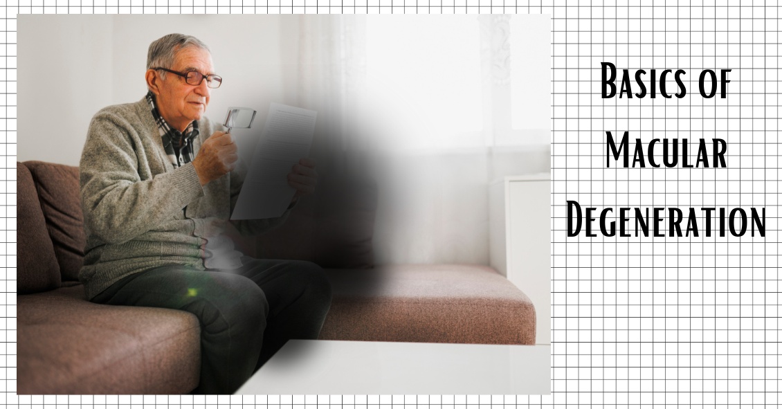
Age-related macular degeneration, often called ARMD or AMD, is the leading cause of vision loss among Americans 65 and older.

AMD causes damage to the macula, which is the central portion of the retina responsible for sharp central vision. AMD doesn't lead to complete blindness because peripheral vision is still intact, but the loss of central vision can interfere with simple everyday activities such as reading and driving, and it can be very debilitating.

Types of Macular Degeneration

There are two types of macular degeneration: Dry AMD and Wet AMD.

Dry (non-exudative) macular degeneration constitutes approximately 85-90% of all cases of AMD. Dry AMD results from thinning of the macula or the deposition of yellow pigment known as drusen in the macula. There may be gradual loss of central vision with dry AMD, but it is usually not as severe as wet AMD vision loss. However, dry AMD can slowly progress to late-stage geographic atrophy, which can cause severe vision loss.

Wet (exudative) macular degeneration makes up the remaining 10-15% of cases. Exudative or neovascular refers to the growth of new blood vessels in the macula, where they are not normally present. The wet form usually leads to more serious vision loss than the dry form.

AMD Risk factors

  • Age is the biggest risk factor. Risk increases with age.
  • Smoking. Research shows that smoking increases your risk.
  • Family history. People with a family history of AMD are at higher risk.
  • Race. AMD is more common in Caucasians than other races, but it exists in every ethnicity.
  • Gender. AMD is more common in women than men.

Detection of AMD

There are several tests that are used to detect AMD.

A dilated eye exam can detect AMD. Once the eyes are dilated, the macula can be viewed by the ophthalmologist or optometrist. The presence of drusen and pigmentary changes can then be detected.

An Amsler Grid test uses pattern of straight lines that resemble a checkerboard. It can be used to monitor changes in vision. The onset of AMD can cause the lines on the grid to disappear or appear wavy and distorted.

Fluorescein Angiogram is a test performed in the office. A fluorescent dye is injected into the arm and then a series of pictures are taken as the dye passes through the circulatory system in the back of the eye.

Optical coherence tomography (OCT) is a test based on ultrasound. It is a painless study where high-resolution pictures are taken of the retina.

Subcategories

Mission Statement

Our doctors and staff are committed to providing thorough care with personal attention. At Eye Doctors of Madison, you will find the compassionate care of a small-town doctors' office with the knowledge of a big-city institution. It is our mission to not only treat each patient uniquely but also like family.

Latest News

Pregnancy and Eye Changes
Pregnancy and Eye Changes We all know that during pregnancy, a woman's body goes through a great deal of change hormonally and physiologically.  But did you know her eyes change as well?  ...
3 Blinding Diseases We Check for During...
3 Blinding Diseases We Check for During an Eye Exam We sometimes get asked, "Why do I need an eye exam when I can see great?" An eye exam doesn't just check your visual acuity--we are also looking...